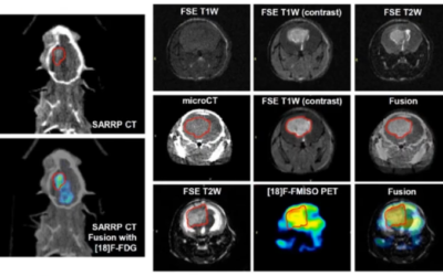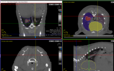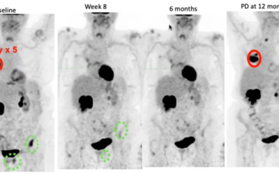
Bioluminescence imaging (BLI) is a non-contact, optical imaging technique based on measurement of emitted light due to an internal source, which is often directly related to cellular activity. It is widely used in pre-clinical small animal imaging studies (such as those preformed on the Xstrahl SARRP) to assess the progression of diseases such as cancer, aiding in the development of new treatments and therapies. For many applications, the quantitative assessment of accurate cellular activity and spatial distribution is desirable as it would enable direct monitoring for prognostic evaluation.
Combined radiotherapy and hyperthermia treatments may improve treatment outcome by heat induced radio-sensitisation.
In their paper “A comprehensive model for heat-induced radio-sensitisation” Brüningk SC, Ijaz J, Rivens I, Nill S, Ter Haar G, Oelfke U, propose an empirical cell survival model (AlphaR model) to describe this multimodality therapy.
The model is motivated by the observation that heat induced radio-sensitisation may be explained by a reduction in the DNA damage repair capacity of heated cells. They assumed that this repair is only possible up to a threshold level above which survival will decrease exponentially with dose. Experimental cell survival data from two cell lines (HCT116, Cal27) were considered along with that taken from the literature (baby hamster kidney and Chinese hamster ovary cells) for hyperthermia and combined radiotherapy-hyperthermia using the Xstrahl SARRP. The AlphaR model was used to study the dependence of clonogenic survival on treatment temperature, and thermal dose R2 ≥ 0.95 for all fits).
For hyperthermia survival curves (0-80 CEM43 at 43.5-57 °C), the number of free fit AlphaR model parameters could be reduced to two. Both parameters increased exponentially with temperature.
They derived the relative biological effectiveness (RBE) or hyperthermia treatments at different temperatures, to provide an alternative description of thermal dose, based on the AlphaR model. For combined radiotherapy-hyperthermia, the analysis is restricted to the linear quadratic arm of the model. They show that, for the range used (20-80 CEM43, 0-12 Gy), thermal dose is a valid indicator of heat induced radio-sensitisation, and that the model parameters can be described as a function thereof. Overall, the proposed model provides a flexible framework for describing cell survival curves, and may contribute to better quantification of heat induced radio-sensitisation, and thermal dose in general.
This Xstrahl In Action was adapted from a article found on a National Library of Medicine website.
SARRP Research Spotlight: Dr. George Wilson
George Wilson, PhD, Chief, Radiation Biology, William Beaumont Hospital Radiation Biology focuses on translational research in the areas of new treatments, combined modalities, and stem cell biology. The group has a heavy emphasis on incorporating molecular,...


