PURPOSE:
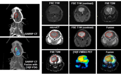
To demonstrate the computed tomography, conformal irradiation, and treatment planning capabilities of a small animal radiation research platform (SARRP).
METHODS AND MATERIALS:
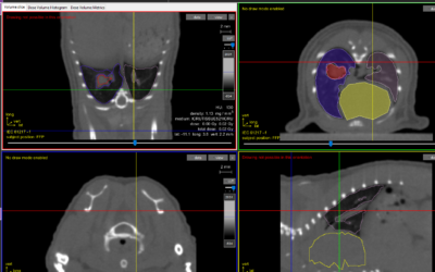
The SARRP uses a dual-focal spot, constant voltage X-ray source mounted on a gantry with a source-to-isocenter distance of 35 cm. Gantry rotation is limited to 120 degrees from vertical. X-rays of 80-100 kVp from the smaller 0.4-mm focal spot are used for imaging. Both 0.4-mm and 3.0-mm focal spots operate at 225 kVp for irradiation. Robotic translate/rotate stages are used to position the animal. Cone-beam computed tomography is achieved by rotating the horizontal animal between the stationary X-ray source and a flat-panel detector. The radiation beams range from 0.5 mm in diameter to 60 x 60 mm(2). Dosimetry is measured with radiochromic films. Monte Carlo dose calculations are used for treatment planning. The combination of gantry and robotic stage motions facilitate conformal irradiation.
RESULTS:
The SARRP spans 3 ft x 4 ft x 6 ft (width x length x height). Depending on the filtration, the isocenter dose outputs at a 1-cm depth in water were 22-375 cGy/min from the smallest to the largest radiation fields. The 20-80% dose falloff spanned 0.16 mm. Cone-beam computed tomography with 0.6 x 0.6 x 0.6 mm(3) voxel resolution was acquired with a dose of <1 cGy. Treatment planning was performed at submillimeter resolution.
CONCLUSION:
The capability of the SARRP to deliver highly focal beams to multiple animal model systems provides new research opportunities that more realistically bridge laboratory research and clinical translation.
Wong J, Armour E, Kazanzides P, Iordachita I, Tryggestad E, Deng H, Matinfar M, Kennedy C, Liu Z, Chan T, Gray O, Verhaegen F, McNutt T, Ford E & DeWeese TL.


