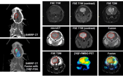
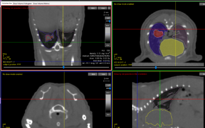
Radiation is used in the study of neurogenesis in the adult mouse both as a model for patients undergoing radiation therapy for CNS malignancies and as a tool to interrupt neurogenesis. We describe the use of a dedicated CT-guided precision device to irradiate specific sub-regions of the adult mouse brain. Improved CT visualization was accomplished with intrathecal injection of iodinated contrast agent, which enhances the lateral ventricles. T2-weighted MRI images were also used for target localization. Visualization of delivered beams (10 Gy) in tissue was accomplished with immunohistochemical staining for the protein γ-H2AX, a marker of DNA double-strand breaks. γ-H2AX stains showed that the lateral ventricle wall could be targeted with an accuracy of 0.19 mm (n = 10). In the hippocampus, γ-H2AX staining showed that the dentate gyrus can be irradiated unilaterally with a localized arc treatment. This resulted in a significant decrease of proliferative neural progenitor cells as measured by Ki-67 staining (P < 0.001) while leaving the contralateral side intact. Two months after localized irradiation, neurogenesis was significantly inhibited in the irradiated region as seen with EdU/NeuN double labeling (P < 0.001). Localized radiation in the rodent brain is a promising new tool for the study of neurogenesis. E. C. Ford, P. Achanta, D. Purger, M. Armour, J. Reyes, J. Fong, L. Kleinberg, K. Redmond, J. Wong, M. H. Jang, H. Jun,d H-J. Song, & A. Quinones-Hinojosab. Download Paper
SARRP Research Spotlight: Dr. George Wilson
George Wilson, PhD, Chief, Radiation Biology, William Beaumont Hospital Radiation Biology focuses on translational research in the areas of new treatments, combined modalities, and stem cell biology. The group has a heavy emphasis on incorporating molecular,...


