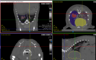
This study examines variations of bone and mucosal doses with variable soft tissue and bone thicknesses, mimicking the oral or nasal cavity in skin radiation therapy. Monte Carlo simulations (EGSnrc-based codes) using the clinical kilovoltage (kVp) photon and megavoltage (MeV) electron beams, and the pencil-beam algorithm (Pinnacle(3) treatment planning system) using the MeV electron beams were performed in dose calculations. Phase-space files for the 105 and 220 kVp beams (Gulmay D3225 x-ray machine), and the 4 and 6 MeV electron beams (Varian 21 EX linear accelerator) with a field size of 5 cm diameter were generated using the BEAMnrc code, and verified using measurements. Inhomogeneous phantoms containing uniform water, bone and air layers were irradiated by the kVp photon and MeV electron beams. Relative depth, bone and mucosal doses were calculated for the uniform water and bone layers which were varied in thickness in the ranges of 0.5-2 cm and 0.2-1 cm. A uniform water layer of bolus with thickness equal to the depth of maximum dose (d(max)) of the electron beams (0.7 cm for 4 MeV and 1.5 cm for 6 MeV) was added on top of the phantom to ensure that the maximum dose was at the phantom surface. From our Monte Carlo results, the 4 and 6 MeV electron beams were found to produce insignificant bone and mucosal dose (<1%), when the uniform water layer at the phantom surface was thicker than 1.5 cm. When considering the 0.5 cm thin uniform water and bone layers, the 4 MeV electron beam deposited less bone and mucosal dose than the 6 MeV beam. Moreover, it was found that the 105 kVp beam produced more than twice the dose to bone than the 220 kVp beam when the uniform water thickness at the phantom surface was small (0.5 cm). However, the difference in bone dose enhancement between the 105 and 220 kVp beams became smaller when the thicknesses of the uniform water and bone layers in the phantom increased. Dose in the second bone layer interfacing with air was found to be higher for the 220 kVp beam than that of the 105 kVp beam, when the bone thickness was 1 cm. In this study, dose deviations of bone and mucosal layers of 18% and 17% were found between our results from Monte Carlo simulation and the pencil-beam algorithm, which overestimated the doses. Relative depth, bone and mucosal doses were studied by varying the beam nature, beam energy and thicknesses of the bone and uniform water using an inhomogeneous phantom to model the oral or nasal cavity. While the dose distribution in the pharynx region is unavailable due to the lack of a commercial treatment planning system commissioned for kVp beam planning in skin radiation therapy, our study provided an essential insight into the radiation staff to justify and estimate bone and mucosal dose. Chow JC, Jiang R. Download Paper
SARRP Research Spotlight: Dr. George Wilson
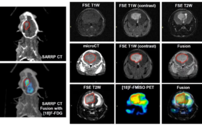
George Wilson, PhD, Chief, Radiation Biology, William Beaumont Hospital Radiation Biology focuses on translational research in the areas of new treatments, combined modalities, and stem cell biology. The group has a heavy emphasis on incorporating molecular,...


