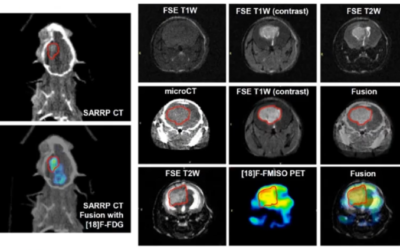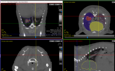
Recently I was asked a question by a colleague, “what is defined by image guided micro-irradiation?” I started to give my normal description along the lines of the process involving imaging, identifying the target in space, being able to target a beam of radiation to a specific area, utilizing treatment planning software, being clinically translational, etc. and so on. But, what I realized during this conversation is that the most important part of the definition, was not that it is image guided. More importantly, it is the notion of identifying the target in space.
Space is defined as the dimensions of height, depth, and width within which all things exist and move. So by definition, if you’re identifying something in space, you need to take into consideration 3 separate dimensions. This is a very important concept to understand when targeting a micro beam of radiation to a target that is located within a small animal. In order to identify the target in space, a 3D image (X, Y, and Z planes) must be acquired. This is commonly achieved through an imaging technique called cone beam-CT, which usually captures X-ray images from at least 360 degrees around the specimen and reconstructs the image into a 3D render. The result is being able to identify the target in space (height, depth, width) because you have performed a 3D imaging technique.
Clinically, the term image guided radiation therapy (IGRT) is understood to incorporate 3D imaging. However, not all pre-clinical imaging combined with radiation delivery is 3D. Therefore, the term “image guided micro-irradiation” (IGMI) can be very misleading. There are plenty of pre-clinical imaging devices that utilize X-ray technology, bioluminescence, fluorescence, and NIR to name a few. Most of them, do not take into consideration height, depth, and width. Performing 3D imaging is a difficult task technologically, so most systems have been designed to give a qualitative 2D image that identifies an area of interest. The actual location of the desired target, if imaged in 2D, can be quite ambiguous as the technique does not capture the target is space. As it stands, the term “image guided” encompasses both 2D and 3D pre-clinical imaging. This is misleading to researchers who aren’t accustomed to the technologies, and can produce confounding or unexpected results due to variables that were expected to be accounted for because the system incorporates “image guidance”.
I think we have all ordered from a catalog and awaited anticipating the arrival of our desired object, only to be disappointed when you open the box. Or, maybe it’s a discussion with a not so transparent sales person who leads you to believe what you desire in a product. But in reality, it’s not exactly what you wanted or the product performs different as expected. Why does this matter in the context of radiation you may ask? It’s simple. If you are not imaging in 3D, then you cannot truly target. The whole concept of “targeting” radiation, is to deliver the most conformal 3D dose to a specific point in space while minimizing exposure to the surrounding normal tissue. How can you pinpoint something in space if you only image in 2D? The answer is, you can’t!
You might be asking yourself, now what? It’s my opinion that the time has come to break the mold on how we, the scientific community refers to these technologies. It’s important we differentiate between the two types of systems that make up IGMIs by imaging capabilities. If you are imaging in a single plane, then you’re performing 2D image guided micro-irradiation (2D IGMI). If you are performing 3D imaging (for instance cone beam-CT), you guessed it, you’re performing 3D image guided micro- irradiation (3D IGMI). I won’t argue with the suffix of the term changing, but the important part is being 100% clear about the imaging. As this will dictate the accuracy and overall reproducibility of your experiments.

This Xstrahl In Action was adapted from a article found on a National Library of Medicine website.
SARRP Research Spotlight: Dr. George Wilson
George Wilson, PhD, Chief, Radiation Biology, William Beaumont Hospital Radiation Biology focuses on translational research in the areas of new treatments, combined modalities, and stem cell biology. The group has a heavy emphasis on incorporating molecular,...


